
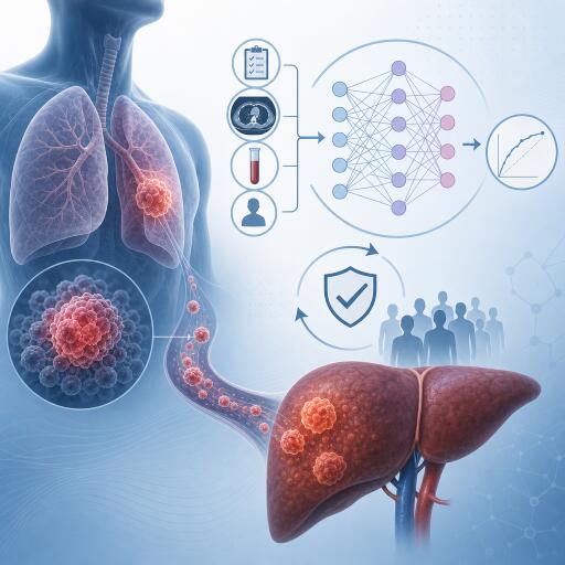

Development and internal validation of a machine learning-based clinical prediction model for liver metastasis in stage III-IV non-small cell lung cancer
A newly accepted study suggests that machine learning could help clinicians identify which patients with advanced non-small cell lung cancer (NSCLC) are at highest risk of developing liver metastasis. The work focuses on stage III-IV disease, where liver involvement is a major driver of poor outcomes and can substantially influence treatment planning.
The researchers analyzed data from 849 patients with stage III-IV NSCLC in a retrospective study. To build and test their model, they split the cohort into two groups: a training set of 679 patients and an internal validation set of 170 patients. Their goal was to create a practical prediction tool that could flag high-risk patients earlier, using routinely available clinical and laboratory data.
To narrow down the most informative variables, the team used a three-part feature selection strategy combining LASSO regression, random forest, and decision tree methods. Rather than relying on a single algorithm, they looked for overlap across all three approaches. That Venn-style intersection produced six shared predictors that consistently stood out: lactate dehydrogenase (LDH), multi-organ metastasis, hemoglobin (HGB), thrombin time (TT), platelet count (PLT), and alkaline phosphatase (ALP).
These markers reflect a mix of tumor burden, blood characteristics, and systemic physiological changes. Elevated LDH is often associated with more aggressive disease, while multi-organ metastasis indicates broader cancer spread. Hemoglobin, thrombin time, platelet count, and alkaline phosphatase may capture additional signals related to anemia, coagulation, inflammation, and liver or bone involvement.
With those six inputs selected, the investigators trained 10 different machine learning classifiers. They then combined two top-performing models with complementary strengths into a Stacking ensemble, a method that aims to improve accuracy by blending multiple algorithms rather than depending on just one.
On the internal validation set, the Stacking model delivered the best overall performance. It achieved an area under the ROC curve (AUC) of 0.852, with a 95% confidence interval of 0.769 to 0.921, indicating strong discriminatory power. It also posted an area under the precision-recall curve (AUPRC) of 0.565, a sensitivity of 76.0%, and a negative predictive value (NPV) of 95.2%.
In practical terms, the model appears especially useful for ruling out liver metastasis risk in many patients, given its high negative predictive value. That could make it a helpful triage tool in oncology workflows, particularly when clinicians need to decide who may benefit from closer imaging surveillance or more aggressive assessment.
The researchers also evaluated calibration, which measures how well predicted probabilities align with real-world outcomes. According to the study, the calibration curves showed that the model’s risk estimates were well matched to observed results. Decision curve analysis further suggested that using the model could provide a net clinical benefit, supporting its potential value in real care settings rather than only in statistical testing.
To make the model more interpretable, the team used SHAP analysis, a popular explainability technique in machine learning. That analysis highlighted elevated LDH, especially around or above 250 U/L, and multi-organ metastasis as the two strongest contributors to prediction. Lower hemoglobin, shortened thrombin time, and abnormal platelet count or alkaline phosphatase also added independent predictive value, though their effects were smaller.
Interpretability matters because clinical AI tools are far more likely to gain traction when physicians can understand what is driving the output. In this case, the model does not depend on obscure genomic features or hard-to-obtain scans alone. Instead, it leans on variables that are generally familiar and accessible in hospital practice.
The authors say they have already deployed an online calculator for point-of-care use, suggesting an emphasis on translational impact rather than purely academic model building. If validated more broadly, such a tool could help oncologists stratify patients with advanced NSCLC and potentially tailor follow-up strategies earlier in the disease course.
Still, there are important caveats. This was a retrospective study, and the validation was internal rather than external. That means the model has so far been tested only within the same overall patient population from which it was developed. Before it can be considered ready for broad clinical adoption, it will need prospective testing and independent validation in outside cohorts.
Even so, the study offers a strong example of how machine learning is moving beyond headline-grabbing experimentation and into focused clinical applications. By targeting a specific, high-impact complication in advanced lung cancer and building on routinely available data, the researchers have created a model that appears both technically solid and potentially practical.
For now, the message is clear: a Stacking-based machine learning approach may provide a reliable way to predict liver metastasis risk in stage III-IV NSCLC, with strong discrimination, solid calibration, and a visible path toward clinical decision support. The next step is proving that performance holds up in real-world settings beyond the original dataset.